Structure and dynamics are the most fundamental concepts of chemistry. Yet, even though we have a fundamental understanding of the nature of the chemical bond and its dynamics, the multitude of interactions present in large systems frequently leads to an overwhelming complexity. For example, predicting the structure of a protein from its amino acid sequence or even designing a protein to perform a specific task are daunting challenges. At the same time, the study of complex systems at different length and timescales promises exciting new insights and discoveries. The goal of capturing complex processes has spurred the development of methods that probe the atomic positions directly and with ultrafast time resolution.
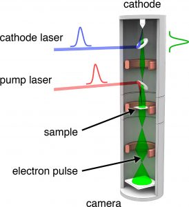
Our method of choice is Time-resolved or Ultrafast Electron Microscopy, which combines the spatial resolution of Electron Microscopes with the time resolution of modern laser systems. The sample is irradiated with a short pump laser pulse to trigger a process. A time-delayed second laser pulse strikes the cathode to create a short electron pulse, which interacts with the sample to capture a snap shot of the dynamics. The instrument thus combines the full scope of established Electron Microscopy techniques with the time resolution of modern laser systems, down to the femtosecond timescale, making it a very powerful tool to study dynamics at different length and timescales.
We are particularly interested in systems whose complex behavior is of a molecular origin and determined by chemical interactions. This is for example frequently the case for nanoscale systems that show specific finite size effects. A macroscopic body is well described by continuum theories. However, as the size of the object is reduced, its macroscopic properties start to disappear, and the molecular properties begin to manifest themselves.
Some recent work in which we studied liquid flow in individual nanotubes may serve to illustrate this point. Whereas the flow of a liquid through a macroscopic tube is well understood, nanoscale flow phenomena may significantly differ from the predictions of continuum theory. While such nanoscale effects are highly intriguing and might be exploited for novel kinds of fluidic devices, their investigation is very challenging, since it requires both high spatial and temporal resolution.
Our experimental approach involved melting the lead core of a ZnO nanotube with a short laser pulse to create a hot, pressurized liquid, whose expansion subsequently drives a number of nanoscale flow phenomena. In particular, when we observed the expansion of a liquid column inside a nanotube, we found that the flow is enhanced by at least one order of magnitude compared with the expectations of macroscopic continuum theory. The origin of this effect appears to a lie in the reduced friction between the liquid and tube wall. Nanoscale viscous friction, however, is a complex interfacial phenomenon, resulting from the dynamical interaction of a large number of atoms or molecules.
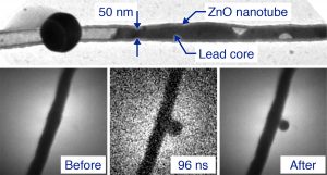
Top: The lead core of a filled ZnO nanotube is melted under irradiation with a series of laser pulses. Bottom: Formation of a liquid extrusion on the side of a nanotube, captured in a single shot image.
U. J. Lorenz, A. H. Zewail,
Science 2014, 344, 1496-1500.
It should also be noted that the possibility to use time-resolved Electron Microscopy to study nanofluidic phenomena is a surprising result, considering that neither traditional Electron Microscopy nor time-resolved laser experiments find widespread application to nanofluidic problems. The marriage of both fields however enables entirely new applications, underlining the power of this rapidly developing technique.